A tiny microscope that may be manoeuvred by means of small areas contained in the physique throughout surgical procedure might pace up breast most cancers remedy and assist lower NHS ready lists, say scientists.
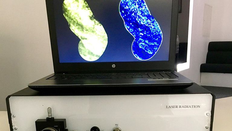
Experts from Imperial College London have developed an endo-microscope that’s lower than 1mm in diameter – in regards to the width of 25 human hairs – and is designed to be inserted into the physique to offer views of tissue and organs.
The machine is ready to produce photos from contained in the tissue with “unprecedented speed” – as much as 120 frames per second, the staff stated.
The hope is the endo-microscope, which is being developed by Dr Khushi Vyas and school colleagues, will assist surgeons establish cancerous cells a hundredth of a millimetre in measurement at a a lot quicker charge than conventional strategies.
This will, the staff says, assist scale back the necessity for follow-up operations to take away cancerous cells that beforehand evaded detection.
Read extra:
Most girls cannot spot signs of ‘sneaky’ type of aggressive breast most cancers, research finds
Cancer vaccine might grow to be out there by 2030, scientists behind COVID jab say
Couch potatoes ‘extra more likely to get breast most cancers,’ consultants warn
The instrument will even assist with breast-conserving surgical procedure, the place the surgeon removes the most cancers whereas leaving as a lot regular breast as attainable.
Up to twenty% of sufferers handled, at the moment want such operations.
The growth of the endo-microscope is being supported by the Engineering and Physical Sciences Research Council (EPSRC), a part of UK Research and Innovation.
‘Available in 5 years’
EPSRC director for cross-council programmes, Dr Kedar Pandya, stated: “By reducing the time it takes to identify cancerous cells and improve the accuracy of imaging, the endo-microscope developed by Dr Vyas and his team could benefit patients and the NHS by reducing waiting lists.”
He added the purpose was to push on with scientific trials with a view to it changing into out there in about 5 years.
The researchers have used their system for preliminary research on human most cancers tissue and are actually testing its use by surgeons and pathologists on laboratory samples of cancerous tissue.
Source: information.sky.com”