
The University of British Columbia (UBC) issued a statement saying, “This picture has been taken at atomic-resolution, which will provide important information as to why the B.1.1.7 variant is more contagious.”

Token photo
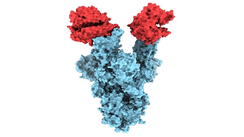
Researchers from Canada have published the first image (B.1.1.7 variant image) of the B.1.1.7 variant of the COVID-19 virus. Through this, it will be known why it is so much more contagious than the previously found variants of the virus. Due to the B.1.1.7 variant, not only the cases of corona increased in the UK, but also in India and Canada it became the cause of increase in cases of infection. The World Health Organization (WHO) reported the first case of the B.1.1.7 variant in mid-December last year. Due to this, a large number of mutations were seen.
The University of British Columbia (UBC) issued a statement saying, “This picture has been taken at atomic-resolution, which will provide important information as to why the B.1.1.7 variant is more contagious.” B.1.1.7 variants were first discovered in Britain. Currently, this variant is the cause of Corona cases coming across Canada. This team of UBC researchers was led by Dr. Sriram Subramaniam. He is a professor at UBC’s Medicine Department of Biochemistry and Molecular Biology Faculty.
First picture of B.1.1.7 variants (University of British Columbia)
Dr. Sriram Subramaniam gave this information regarding the B.1.1.7 variant
Dr. Sriram Subramaniam was particularly interested in a mutation called N501Y found in the spike protein of coronavirus. The coronavirus attaches itself to the cells present in the human body and infects it. He said, the pictures taken by us show the first structural glimpse of the N501Y mutant. It also shows that the changes occurring in it happen locally.
Dr. Subramaniam stated that the N501Y mutation is actually the only mutation in the B.1.1.7 variant that is located in the spike protein. This is what binds the ACE2 receptor present in the human body. The ACE2 receptor is an enzyme present on the surface of our body’s cells, which works by entry to the Sars-CoV-2 virus.
Photograph taken through a special microscope
The coronavirus is up to a million times smaller than the pin point and it is extremely difficult to detect with a normal microscope. The research team used a ‘cryo-electron microscope’, also known as cryo-EM, to detect the wide size of viruses and proteins.
This microscope is up to 12 feet high and uses electron waves at liquid nitrogen temperature to take pictures. Dr. Subramaniam stated that the photograph taken through our cryo-EM shows the relationship of Y residue (501Y) mutations with ACE2. Regarding this, we believe that this is the reason for increasing the binding and infectivity of B.1.1.7.
Also read: You know how the Corona vaccine is made! Reaches us through 18 difficult stages
.